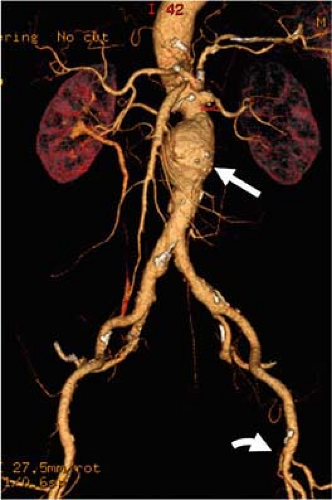
Courtesy to: Abdominal Aorta W. Dennis Foley,F. Scott Pereles
You have a case of 72 years old gentleman presented with Juxta-renal aneurysm 6.7cm
With history of diabetes, hypertension, ischemic heart disease and previous CABG 6 years ago and stable history
After preliminary investigations the plan was to go for 4 fenestrations EVAR through femoral access
Q1: What are the risks to be included in the consent form?
Q2: What are the most important points to look at during planning and arranging the accessories for the case?
Q3: What are the key steps of the procedure from going through the access to the final check angiogram please?
9 Comments
Ahmed Bedeer
Q1: Risks to Include in the Consent Form
FEVAR risks …….
Procedural risks:
Vascular injury
Endoleak
Graft migration or occlusion
Spinal cord ischemia
Renal complications: Injury to the kidneys from contrast dye (contrast-induced nephropathy) or from covering the renal arteries with the graft.
Patient-specific risks:
Cardiac complications: a prior CABG (coronary artery bypass graft), there’s a risk
Renal failure
Cerebral vascular events
Infection
General risks:
Bleeding or hematoma
Allergic reaction
Death
############
Q2: Key Planning and Accessory Points
patient age and comorbidities
aneurysm morphology: the aneurysm’s length, diameter, and angulation.
Anatomical considerations >>Renal and mesenteric artery ostia: the location, diameter, and angle of the renal arteries and superior mesenteric artery (SMA) to ensure the fenestrations on the custom graft align perfectly. >> Proximal and distal landing zones
Access vessels: large enough and not too tortuous for the delivery system.
Tools availability >>> Custom-made fenestrated stent graft: holes (fenestrations) or scallops + Sheaths and delivery systems + Bridging stents + Wires and catheters + Inflation devices and balloons + Contrast injector
###############
Q3: Key Steps of the Procedure
The procedure is a multi-step process that requires precision and meticulous execution for 10 steps of planning……
Centerline placement for accurate measuring
marking the center of visceral artery origen
Determining tha proximal landing zones site with a minimum of 20 mm parallel walled arterial segment free for mural disease
Measuring the diameter of landing zones at 3 sites and the largest of them to be oversized bt 10_15%
Measuring the length from the proximal edge of the graft to the center of each branch vessel
Measuring target artery clock orientation
choose the proximal fenestrated component
indicted the desine scalloping or fenestrations
Choose the distal bifurcated component
choose the iliac limb
And 5 steps of applying…….
Access: The procedure begins with gaining femoral artery access. Typically, both femoral arteries are accessed percutaneously or via a small incision.
Guide wire Placement: A guidewire is advanced from the femoral artery, up into the aorta, and positioned in a safe location, often in the ascending aorta. This acts as a rail for the delivery system.
Graft Deployment: The main fenestrated stent graft is deployed. The delivery system is carefully navigated to the correct position, and the graft is released. The key here is to align the fenestrations of the graft with the patient’s renal and SMA arteries. This often involves specific markers on the graft and fluoroscopy.
Bridging Stent Cannulation and Deployment: Once the main graft is in place, the renal and SMA arteries are cannulated. A catheter and wire are used to navigate through the fenestrations into each target vessel. Bridging stents are then deployed within these vessels, extending from the main graft into the target arteries. This secures the connection and prevents leaks.
Final Check Angiogram: After all components are in place, a final angiogram is performed. This involves injecting contrast dye and taking images to confirm that the stent graft is correctly positioned, the visceral arteries are patent, and there are no endoleaks or other complications. If any issues are detected, further ballooning or stent placement may be necessary.
A1
UP to death
MI
Bowel ischemia
Spinal cord ischemia
Endo leak
…..
A2
Age
Aneurysm size
Comorbiditis
Anatomical concidriations
Acesses
…….
A3
Central line into aorta
Place marker for each vesral artery, like renal and SMA
Proximal landing zone of graft is over sizing by 10%
The clock face position of each target artery chose the proximal fenestrated
The distal biforcated body
Iliac limbs
A1 risks
death
MI -AKI -bowel ischemia -spinal cord ischemia -ll ischemia
stroke – acsess complications like pseudoaneurysm and hematoma
endoleak
A2 factors
patient age
comorbidities
aneurysm size
anatomical consideration and viratation
acsess
proximal and distal landing zone
A3 key steps
starts with central line placement into the aorta
place markers at the origin of each visceral artery — like renals, SMA
the proximal landing zone of the graft. with oversizing the graft by about 10%.
the clock-face position of each target artery
choose the proximal fenestrated component,
the distal bifurcated body, and finally the iliac limbs.
Q1:
Complications can include MI, renal failure, or stroke. Also things like endoleak, limb thrombosis, or stent migration. On top of that, there’s risk of wound infection and even secondary hemorrhage post-op.
⸻
Q2:
The decision depends on multiple factors — patient’s age, size of the aneurysm, his comorbidities like HTN, DM, IHD, etc. Also anatomical factors like neck length, tortuosity, and whether the access vessels are suitable or not.
⸻
Q3:
It starts with central line placement into the aorta, then we place markers at the origin of each visceral artery — like renals, SMA — and also at the proximal landing zone of the graft. We usually oversize the graft by about 10%. Need to know the clock-face position of each target artery, then choose the proximal fenestrated component, the distal bifurcated body, and finally the iliac limbs.
Q1:
MI, Renal failure, stroke
endoleak, limb thrombosis or stent migration
wound infection and secondary hemorrahge
Q2:
age of the patient and aneurysmal size and his medical comorbidities and anatomical considerations and the access vessel requirements
Q3:
10 steps central line placement of the aorta and marker placement in the center of each of the visceral artery origin and proximal end of the device and 10% oversizing and target artery clock position and choose the proximal fenestration component and distal bifurcated component and iliac limb
A1..
Intra operative type 1A or B endo leak
Early post op complications
*.Access sites complications as hematoma and pseudoaneurysm or wound infection
Early graft thrombosis
*. General cardiac and respiratory complication
Ranal failure
Late complications as
Endoleake . Graft migration or thrombosis
Q2..
> access sites (diameter ,torsousity,calcification and thrombosis)
> Landing zones at least 20mm healthy parallel wall of arterial segment free from thrombus of calcification
> Clock Orientation of branch vessels and their diameters
Q3.
10 steps of planning.
1. Centerline placement for accurate measuring
2.marking the center of visceral artery origen
3. Determining tha proximal landing zones site with a minimum of 20 mm parallel walled arterial segment free for m disease
4. Measuring the diameter of landing zones at 3 sites and the largest of them to be oversized bt 10_15%
5. Measuring the length from the proximal edge of the graft to the center of each branch vessel
6. Measuring target artery clock orientation
7.. chose the proximal fenestrated component
8 indicted the desine scalloping or fenestrations
9 . Choose the distal bifurcated component
10.. choosing the iliac climb
A1:
General MI RF Resp F Stroke DVT
Local Early
Endoleak thrombosis limb occlusion rupture colonic limb renal ischemia CIN access site complication
Local late
Infection migration delayed endoleak endotension
A2:
Patient selection age comorbidities
Anatomical considerations the neck proximal landing zone at least 2 cm parallel disease free and the distal landing zone along with the access site and the whole aorta as well with branch clock orientation for good accurate planning
A3:
10steps for the FEVAR planning
Central line placement
Marking og each br origin
Prox landing zone
Measurements of diameters select tha largest of 3 with 10-15% oversizing
The same for lengths from PLZ to each br origin and to bifurcation
Target vessel clockwise orientation
Choose the proximal fenestsration component and decide if scalop or fenestration along with the bifurcated body graft and iliac limbs
Q1: Risks to Include in the Consent Form
FEVAR risks …….
Procedural risks:
Patient-specific risks:
General risks:
############
Q2: Key Planning and Accessory Points
###############
Q3: Key Steps of the Procedure
The procedure is a multi-step process that requires precision and meticulous execution for 10 steps of planning……
And 5 steps of applying…….
A1
UP to death
MI
Bowel ischemia
Spinal cord ischemia
Endo leak
…..
A2
Age
Aneurysm size
Comorbiditis
Anatomical concidriations
Acesses
…….
A3
Central line into aorta
Place marker for each vesral artery, like renal and SMA
Proximal landing zone of graft is over sizing by 10%
The clock face position of each target artery chose the proximal fenestrated
The distal biforcated body
Iliac limbs
A1 risks
death
MI -AKI -bowel ischemia -spinal cord ischemia -ll ischemia
stroke – acsess complications like pseudoaneurysm and hematoma
endoleak
A2 factors
patient age
comorbidities
aneurysm size
anatomical consideration and viratation
acsess
proximal and distal landing zone
A3 key steps
starts with central line placement into the aorta
place markers at the origin of each visceral artery — like renals, SMA
the proximal landing zone of the graft. with oversizing the graft by about 10%.
the clock-face position of each target artery
choose the proximal fenestrated component,
the distal bifurcated body, and finally the iliac limbs.
Q1:
Complications can include MI, renal failure, or stroke. Also things like endoleak, limb thrombosis, or stent migration. On top of that, there’s risk of wound infection and even secondary hemorrhage post-op.
⸻
Q2:
The decision depends on multiple factors — patient’s age, size of the aneurysm, his comorbidities like HTN, DM, IHD, etc. Also anatomical factors like neck length, tortuosity, and whether the access vessels are suitable or not.
⸻
Q3:
It starts with central line placement into the aorta, then we place markers at the origin of each visceral artery — like renals, SMA — and also at the proximal landing zone of the graft. We usually oversize the graft by about 10%. Need to know the clock-face position of each target artery, then choose the proximal fenestrated component, the distal bifurcated body, and finally the iliac limbs.
Q1:
MI, Renal failure, stroke
endoleak, limb thrombosis or stent migration
wound infection and secondary hemorrahge
Q2:
age of the patient and aneurysmal size and his medical comorbidities and anatomical considerations and the access vessel requirements
Q3:
10 steps central line placement of the aorta and marker placement in the center of each of the visceral artery origin and proximal end of the device and 10% oversizing and target artery clock position and choose the proximal fenestration component and distal bifurcated component and iliac limb
A1..
Intra operative type 1A or B endo leak
Early post op complications
*.Access sites complications as hematoma and pseudoaneurysm or wound infection
Early graft thrombosis
*. General cardiac and respiratory complication
Ranal failure
Late complications as
Endoleake . Graft migration or thrombosis
Q2..
> access sites (diameter ,torsousity,calcification and thrombosis)
> Landing zones at least 20mm healthy parallel wall of arterial segment free from thrombus of calcification
> Clock Orientation of branch vessels and their diameters
Q3.
10 steps of planning.
1. Centerline placement for accurate measuring
2.marking the center of visceral artery origen
3. Determining tha proximal landing zones site with a minimum of 20 mm parallel walled arterial segment free for m disease
4. Measuring the diameter of landing zones at 3 sites and the largest of them to be oversized bt 10_15%
5. Measuring the length from the proximal edge of the graft to the center of each branch vessel
6. Measuring target artery clock orientation
7.. chose the proximal fenestrated component
8 indicted the desine scalloping or fenestrations
9 . Choose the distal bifurcated component
10.. choosing the iliac climb
Thanks a lot for your answer
I would add bowel schema and spinal cord schema to the mentioned possible complications
A1:
General MI RF Resp F Stroke DVT
Local Early
Endoleak thrombosis limb occlusion rupture colonic limb renal ischemia CIN access site complication
Local late
Infection migration delayed endoleak endotension
A2:
Patient selection age comorbidities
Anatomical considerations the neck proximal landing zone at least 2 cm parallel disease free and the distal landing zone along with the access site and the whole aorta as well with branch clock orientation for good accurate planning
A3:
10steps for the FEVAR planning
Central line placement
Marking og each br origin
Prox landing zone
Measurements of diameters select tha largest of 3 with 10-15% oversizing
The same for lengths from PLZ to each br origin and to bifurcation
Target vessel clockwise orientation
Choose the proximal fenestsration component and decide if scalop or fenestration along with the bifurcated body graft and iliac limbs
Well done
very thorough method